
Cartilage Damage: Symptoms and Treatment Options?
Caviarlieri | Published December 22, 2020
Are you suffering from damaged cartilage with distressing symptoms including chronic joint pain, inflammation, a decreased range of movement in the affected joint etc.?
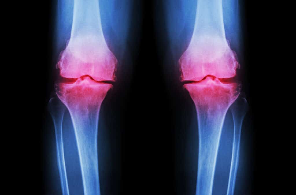
Cartilage covers the surface of joints, enabling bones to slide over one another while reducing friction and preventing damage. It helps to support your weight when you move, bend, stretch and run. Cartilage damage is a relatively common type of injury. It often affects the knees, although joints such as the hips, ankles and elbows also susceptible to cartilage damage.
The Articular cartilage is a complex and specialized tissue that provides a slick and bouncy cushion between the bones at the joints. When this cartilage is damaged by trauma, disease or simple wear and tear, the bones can rub directly against each other, causing pain and inflammation, which can eventually result in arthritis.
How Prevalent is Cartilage Damage?
Loss of this slippery and shock-absorbing tissue layer, called articular cartilage, is responsible for many cases of joint pain and arthritis, which afflicts more than 55 million Americans. Nearly 1 in 4 adult Americans suffer from arthritis but far more are burdened by joint pain and inflammation. In the UK, around 10,000 people yearly have cartilage damage serious enough to warrant treatment.
Cases of accidental cartilage damage are most common in people under 35 years old. This is because this age group is more likely to take part in sporting activities where there is a higher risk of injury. Cartilage damage associated with osteoarthritis is more common in adults over 50 years old. It is also more common in women than in men.
If the damage is severe, a piece of cartilage can break off and become loose. If this happens, the loose piece of cartilage may affect the movement of your joint. This can cause a feeling of the joint ‘locking’ or catching. Sometimes, the joint may also give way, causing serious injury.
The most common location for cartilage damage is in the knee joint. In some cases of knee joint damage, bleeding can occur inside the knee joint. This is known as hemarthrosis and can lead to skin around the joint swelling up.
Treatment Options for Cartilage Damage
The current medical view is that cartilage does not regrow or replace itself, it can only be repaired or supplanted by a few different treatment options. Many cartilage injuries can be treated without surgery, via physical therapy and anti-inflammatory medication. This is especially true if you lead a sedentary to moderately active lifestyle.
1. Non-Surgical Treatment for Cartilage Damage
There are a number of non-surgical treatments that can help relieve symptoms of damaged articular cartilage.
- Physiotherapy – exercises that strengthen the muscles surrounding or supporting your joint which may help reduce pressure on the joint and reduce pain.
- Painkillers – non-steroidal anti-inflammatory drugs (NSAIDs), such as ibuprofen, can help reduce swelling and pain. However, the recommendation is to avoid taking ibuprofen if you have or have had stomach problems, such as stomach ulcer.
- Lifestyle changes – such as reducing activity that involves the affected joint.
In more severe cases of articular cartilage damage, non-surgical treatment may only provide short-term relief and eventually surgery may be required.
2. Surgical Treatment for Cartilage Damage
Surgical treatment for damaged articular cartilage includes the following procedures:
Arthroscopic lavage and debridement
The surgeon makes a cut in the joint before using an arthroscope (a flexible tube with a camera on the end) to assess the damage. They then ‘clean out’ the joint using a saline (salt) solution. Loose cartilage fragments are removed using a device known as a shaver, which works in a similar way to a vacuum cleaner.
Drawback: Arthroscopic lavage and debridement cannot repair the damaged cartilage, but it can help reduce pain and increase mobility.
Marrow stimulation
Marrow stimulation involves making tiny holes (microfractures) into the bone beneath the damaged cartilage using a small pointed instrument known as an awl. This releases the bone marrow from inside the bone and leads to a blood clot forming within the damaged cartilage. The marrow cells then begin to stimulate production of new cartilage.
Drawback: The newly generated cartilage is fibrocartilage rather than hyaline cartilage. As fibrocartilage is not as supple as hyaline cartilage, there is a risk that after a few years it will wear away and further surgery may be needed.
Mosaicplasty
Mosaicplasty is a technique where small rods of healthy cartilage from the non-weight-bearing areas of a joint, such as the side of the knee, are removed and used to replace the damaged cartilage.
Drawback: It is only suitable for treating relatively minor cartilage injury. This is because removing too much healthy cartilage could damage the section of the body from where the cartilage was taken.
Osteotomy
The alignment of the leg is altered slightly to reduce pressure on the damaged area and helps alleviate pain; this usually involves adding or removing a wedge of bone from the shin or thigh and the bone is fixed with a plate until it heals.
Joint Replacement
Replacing the whole joint with an artificial joint, such as a knee replacement or hip replacement is occasionally necessary if the damage is particularly severe.
Allograft Osteochondral Transplantation (AOT)
AOT is a similar procedure to mosaicplasty, but the cartilage is obtained from a recently deceased donor. The cartilage will be tested in a laboratory to make sure it is free from infection before being prepared for transplant.
Autologous Chondrocyte Implantation (ACI)
ACI involves a two-stage technique. During the first stage the surgeon takes a small sample of cartilage cells from the edge of the joint. The cells are then sent to a laboratory and placed in an incubator where they are given nutrients to encourage them to divide and produce new cells. After a few weeks, the number of cartilage cells will have increased by 50 times. The new cartilage cells are used to replace the damaged cartilage.
The second stage involves placing these cells on a collagen patch, which is then sutured or glued onto the damaged area usually through a small incision.
Drawback: After studying ACI, the National Institute for Health and Clinical Excellence (NICE) has decided there is not enough evidence about the procedure’s long-term effects or safety. Therefore ACI is not available on the NHS unless as part of a clinical trial (a type of research that tests one treatment against another).
Cartilage Regeneration
It is evident that none of the current medical treatment options offer cartilage regeneration.
Unlike other types of tissue, the cartilage does not have its own blood supply. Blood cells help to repair damaged tissue, therefore damaged cartilage does not heal as quickly as damaged skin or muscles. Therefore, it is often assumed that humans cannot regrow cartilage tissue, e.g. to reverse the damage of osteoarthritis.
What about PRP and stem cell therapy?
Two options which claimed promise for the successful treatment of cartilage injuries are platelet-rich plasma (PRP) and stem cell therapies. These therapies harness the body’s own immune system to trigger the cartilage to heal and regrow itself in a way it might not otherwise do.
However, these therapies lack scientific evidence and are unproven and is considered by rheumatologists and orthopaedic providers as a grey area of regenerative medicine.
Rejuvenate your Cartilage Before it is Badly Degraded – Try Caviarlieri

Joint replacement surgery has revolutionized how doctors treat arthritis and has become very common: By age 80, 1 in 10 people will have a hip replacement and 1 in 20 will have had a knee replacement. But such joint replacement is extremely invasive, has a limited lifespan and is performed only after the onset of arthritis and patients would have to endure lasting pain. It may be more practical and functional if people are able to avoid getting arthritis in the first place or delay the progression of cartilage degeneration by rejuvenating the cartilage in their joints before it is badly degraded.
Cartilage regeneration, along with strengthening the muscles around the joint, can help some patients delay joint replacement surgery for damaged joints.
Why Caviarlieri – Swiss Caviar Cellular Therapy Supplement?
Caviarlieri is scientifically proven to have very strong anti-inflammatory properties. Formulated with the finest Caviar Cellular Extracts, marine peptides and highly polymerized collagen, it can help reduce chronic inflammation by reducing the Tumour Necrosis Factor-alpha and Hs-C Reactive Protein (inflammatory biomarkers), associated with a broad spectrum of degenerative diseases.
Unlike drugs, steroids and painkillers, which have adverse side effects if used for a long period of time. Caviarlieri is an excellent nutritional supplement which has no side effects and is a great palliative for chronic pain management.
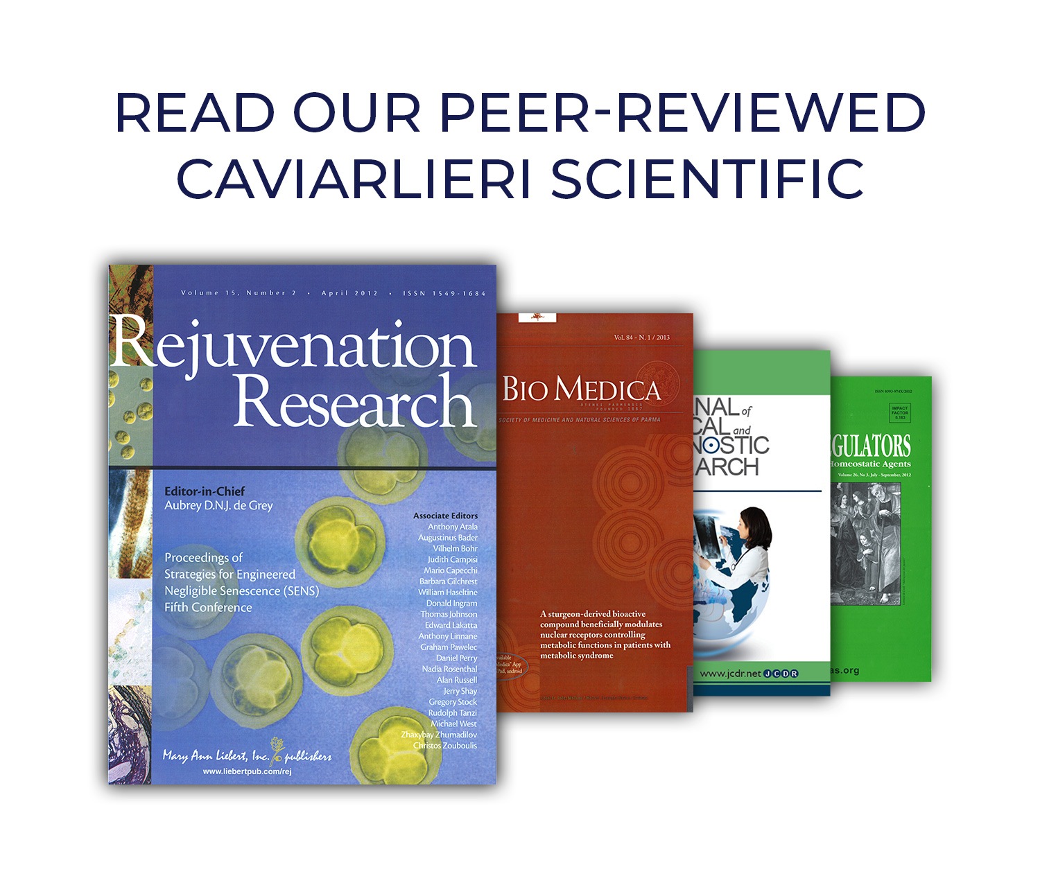
Scientific studies of Caviarlieri published in peer reviewed journals have demonstrated that its primary bio active ingredient, Caviar DNA Extract can trigger the epigenetic mechanisms of the cells. This then activates the gene expression for inflammation reduction and as a result significantly reduces joint pains and inhibit cartilage degradation.
Scientific Evidence
Published in Peer Reviewed Journal – Acta Bio Medical / Official Journal of the Society of Medicine and Natural Sciences of Parma Vol 26, No. 3, July-Sep 2012
BENEFICIAL EFFECT OF A STURGEON-BASED BIOACTIVE COMPOUND ON GENE EXPRESSION OF TUMOR NECROSIS FACTOR-α, MATRIX METALLOPROTEINASES AND TYPE-10 COLLAGEN IN HUMAN CHONDROCYTES
This study examined a possible inhibitory effect of Caviarlieri – Caviar DNA extract (LD-1227) collagen elastin and protein extracts from sturgeon on ILβ -induced activation and production of TNFα and MMP-13 in human osteoarthritis (OA) chondrocytes and intracellular signalling factors. Human chondrocytes were derived from OA cartilage and stimulated with ILβ.
Results
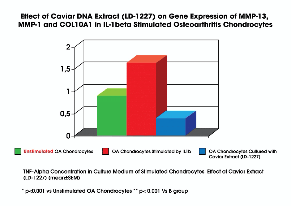
The data showed in the following figures indicate that Caviar DNA extract (LD-1227) can inhibit IL-1β-induced proliferation and inflammatory reactions via inhibited activation of the transcription factor NF-κB pathway in human chondrocytes derived from OA patients. These novel pharmacological actions of Caviarlieri on ILβ-stimulated human OA chondrocytes provide evidence that Caviar DNA extract (LD-1227) may inhibit cartilage degradation by suppressing ILβ-mediated activation and the catabolic response in human chondrocytes.
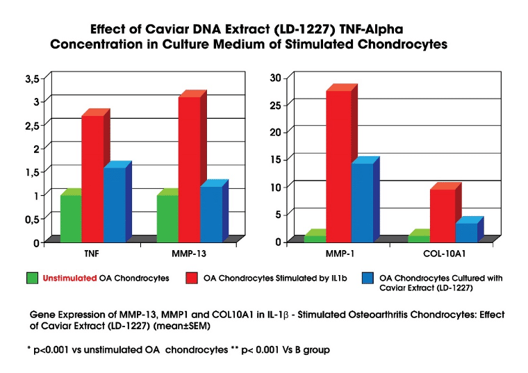
It is evident from the above results that OA chondrocytes (cartilage) cultured with Caviar DNA Extract can significantly lower levels of inflammatory markers like Tumour Necrosis Factor-Alpha, MMP-13, MMP-1 and Collagen type 10. This means that the inflammation markers present in the OA chondrocytes (cartilage) (that were stimulated by IL1b) were significantly reduced when cultured with Caviar DNA Extract (LD-1227).
IMPORTANT: Matrix metalloproteinase 13 (MMP-13) degrades collagenous extracellular matrix and its aberrant activity is associated with diseases such as arthritis, cancer, atherosclerosis and fibrosis.
Note: It is well established that IL-1β stimulates cartilage breakdown. These studies have highlighted the pro-catabolic effects of IL-1 by measuring the expression of inflammatory markers such as MMP levels and cyclooxygenase (COX) and the release of matrix degradation products.
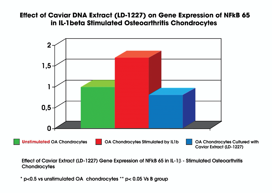
It is evident from the above figure that Caviar DNA Extract (LD-1227) suppresses NF-kB activation. The anti-inflammatory potency of Caviarlieri. Is demonstrated by the suppression of NF-kB activation.
Note: NF-κB induces the expression of various pro-inflammatory genes, including those encoding cytokines and chemokines, and also participates in inflammasome regulation.
Conclusion
Osteoarthritis tends to evolve from the local inflammatory disease of articular cartilage to progressive degenerative joint disease with inflammation and degeneration that ultimately results in underlying bone, pain and disability.
From the data obtained in the study, almost complete inhibition of both TNFα and MMPs expression and production was observed at a concentration of 100 μM Caviar DNA Extract -LD-1227 (p < 0.01), demonstrating that Caviarlieri is a promising new approach for the inhibition of cartilage degradation and more importantly it is scientifically proven to have strong anti-inflammatory properties.


Subject
Recent Posts
-
The Amazing Benefits of Highly Polymerized Fish Collagen Peptides with Elastin – Caviarlieri
-
The World’s Most Effective Caviar DNA Extract with Marine Bioactive Peptides
-
Are Supplements effective for Joint Pain?
-
Why Is Sustainable Immunity Important for Your Long-Term Health
-
What is your “Body Age” – Biological Age?